
What Is the Best Treatment for Receding Gums?
Key Takeaways
Gum recession is permanent—gingival tissue does not regenerate naturally once lost.
The most common causes include periodontal disease, aggressive brushing, thin tissue biotype, and orthodontic movement outside the bone envelope.
The best receding gums treatment depends on severity, cause, tissue type, and esthetic goals—no single solution fits every patient.
Connective tissue grafting (CTG) is the gold-standard treatment for moderate to severe recession, with root coverage predictability exceeding 90% in qualified cases.
The Pinhole Surgical Technique (PST) is a minimally invasive alternative for select patients but carries lower root coverage predictability than CTG in independent studies.
Untreated gum recession leads to root decay, tooth sensitivity, mobility, and eventual tooth loss.
Not all recession defects are fully correctable—Cairo Class III and Miller Class IV defects have guarded prognoses for complete root coverage.
Choosing a board-certified periodontist with advanced soft tissue grafting training is essential for optimal outcomes.
New York City patients experiencing receding gums should seek evaluation from a gum recession specialist, not a general dentist.
Early intervention produces better long-term results—do not wait until recession is severe before seeking treatment.
Why Receding Gums Demand Attention
Gum recession is one of the most underdiagnosed and undertreated conditions in dentistry. Patients often come to my office in Midtown Manhattan after years of noticing a tooth that looks "longer" than the others, only to discover that a significant amount of their protective gingival tissue has been lost—tissue that will not come back on its own.
According to data from the National Health and Nutrition Examination Survey (NHANES), gum recession affects more than 50% of U.S. adults over the age of 30, with prevalence rising steeply with age. Yet despite its commonality, recession is routinely dismissed by patients and, unfortunately, sometimes by clinicians who do not specialize in periodontal tissue management.
What makes gum recession particularly insidious is its progression. It does not happen overnight. It develops slowly, often painlessly, over months or years—until one day a patient notices sensitivity to cold beverages, a darkening or yellowing at the gumline, or an unmistakably elongated appearance to their teeth. By that point, meaningful tissue has already been lost.
Left untreated, receding gums cause a cascade of problems: persistent tooth sensitivity that interferes with daily life, root surface exposure that dramatically increases the risk of root caries (decay on the root surface, which is far more destructive and harder to treat than coronal decay), progressive tooth mobility, esthetic deterioration, and ultimately, tooth loss.
The question I am most often asked is: What is the best treatment for receding gums? The answer is nuanced. The best treatment for receding gums depends on the cause, severity, tissue thickness, esthetic concerns, and long-term prognosis of the affected tooth or teeth. There is no one-size-fits-all solution. However, for most patients presenting with moderate to severe recession defects, connective tissue grafting remains the gold standard—the most predictable, most extensively studied, and most durable treatment available.
This article is written to give patients a comprehensive, honest, and clinically grounded understanding of every treatment option available for gum recession, and to help them make an informed decision about who should treat them and why.
What Are Receding Gums?
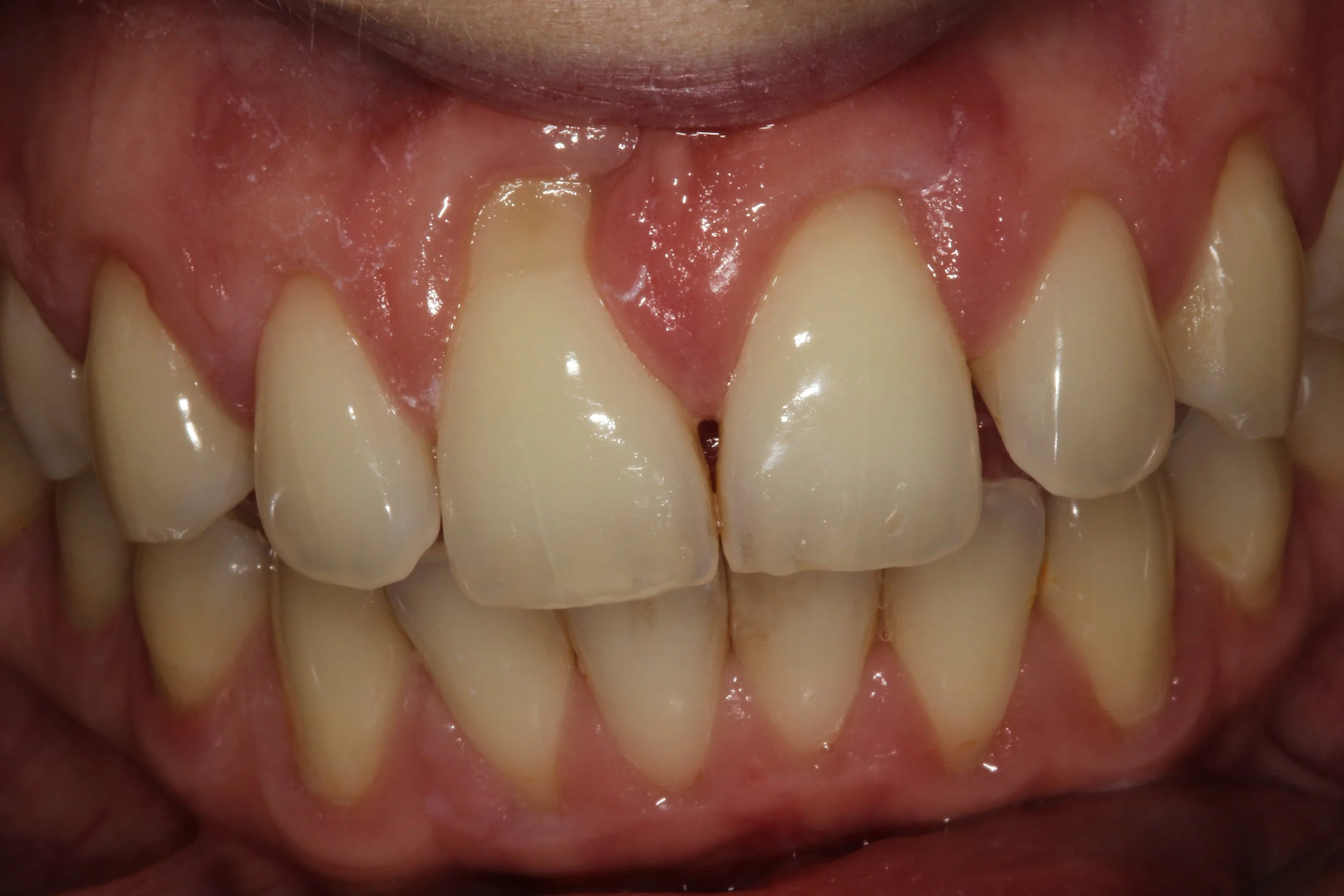
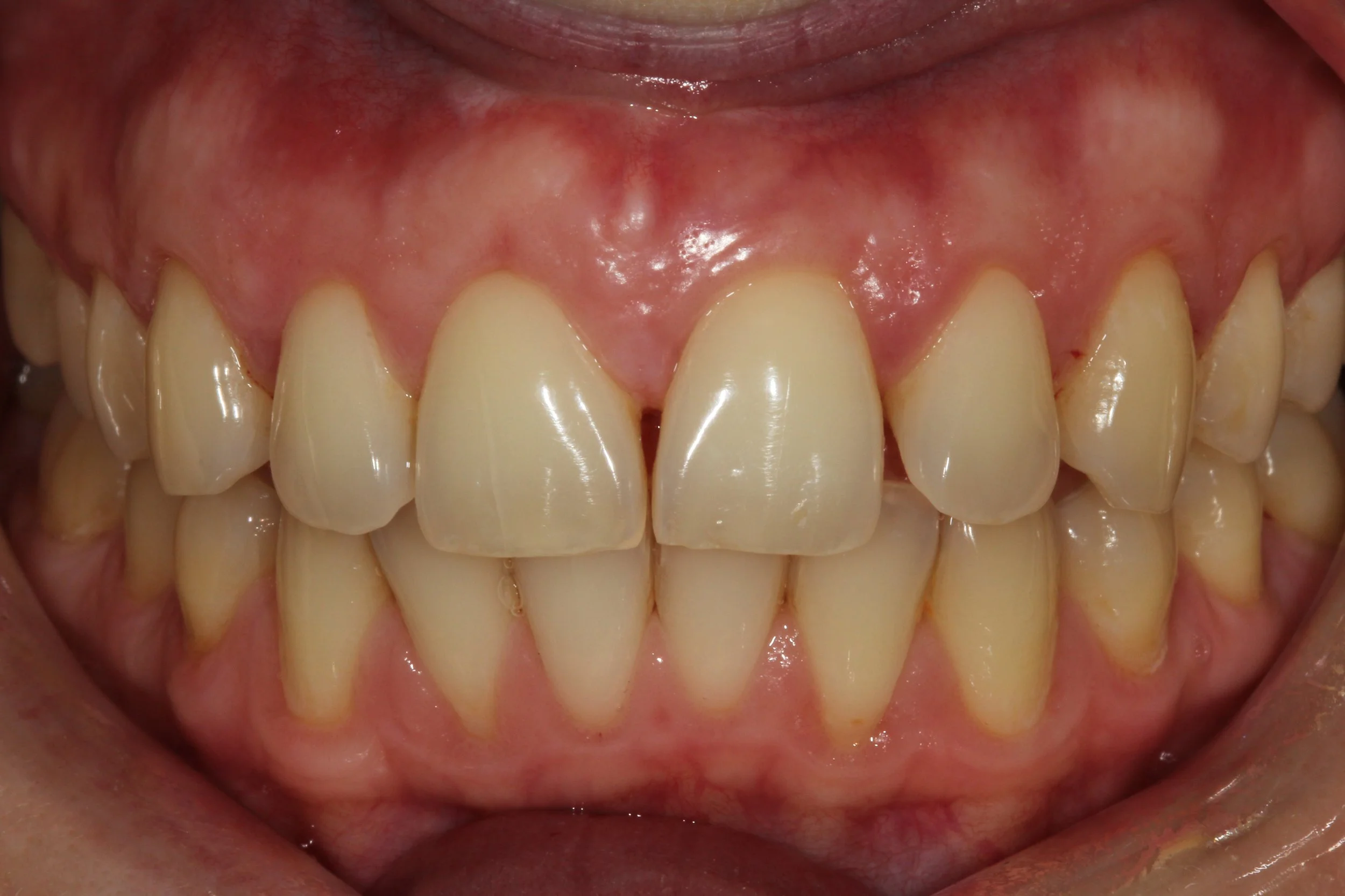
Gum recession, clinically referred to as gingival recession, is the apical migration of the gingival margin away from the cementoenamel junction (CEJ)—the anatomical boundary where the tooth crown meets the root. When recession occurs, the root surface becomes exposed, losing the protective coverage of keratinized gingival tissue.
Healthy gingiva appears pink, firm, stippled, and tightly adapted to the tooth surface. In recession, the tissue margin has migrated downward (on upper teeth) or upward (on lower teeth), exposing the yellowish, softer cementum of the root. The exposed root is not designed to withstand the oral environment the way enamel is: it is susceptible to abrasion, sensitivity, chemical erosion, and bacterial colonization.
There are two distinct tissue types that can be lost in recession:
Attached gingiva: Dense, keratinized tissue firmly bound to the underlying bone and tooth root. This tissue is critical for protecting the tooth from external forces and microbial invasion.
Free gingiva: The non-attached portion of gingiva forming the gingival sulcus. This tissue is less protective but still contributes to the barrier function at the gumline.
Patients with a thin gingival phenotype—characterized by translucent, delicate tissue with minimal keratinized width—are particularly vulnerable to recession. This thin biotype is a genetic characteristic and not something patients can control. In contrast, patients with a thick, fibrous phenotype are more resilient to recession-triggering forces.
Clinically important: recession rarely involves one tooth in isolation. It often appears across multiple adjacent teeth and tends to worsen over time without intervention. The natural history of untreated recession is progressive—it does not stabilize on its own.
How Common Is Gum Recession?
Gum recession is far more prevalent than most patients realize. Epidemiological data consistently demonstrates that it affects the majority of adults, with increasing prevalence as patients age:
Adults aged 30–39: recession prevalence approximately 38%
Adults aged 50–59: prevalence approaching 71%
Adults over 65: prevalence exceeding 88%
However, recession is not exclusively an older adult problem. I regularly treat younger patients—including those in their twenties and early thirties—with clinically significant recession. Several populations warrant special attention:
Athletes: Contact sport athletes, cyclists, triathletes, and patients who train with mouth guards are at elevated risk. Habitual dehydration, acidic sports beverages, and mechanical trauma from equipment all contribute. Athletes also tend to brush vigorously post-workout.
Orthodontic patients: Teeth moved labially (toward the lips) or outside the alveolar bone envelope during orthodontic treatment can develop recession during or after treatment. This is particularly relevant in patients who lacked adequate bone housing prior to treatment—a factor that must be assessed before orthodontic movement begins.
Young adults with aggressive brushing habits: This is one of the most preventable causes of recession. Vigorous brushing with a medium or hard-bristled brush is a leading cause of recession in patients under 40.
Tobacco users: Smokers and smokeless tobacco users develop recession at significantly higher rates than non-users, driven by vasoconstriction, impaired immune response, and direct tissue irritation.
From a public health perspective, the undertreatment of recession is significant. Many patients are told to "watch it" for years by general practitioners who lack the surgical training to intervene. By the time these patients reach a periodontist, simple defects have become complex, multi-tooth reconstruction cases.
What Causes Receding Gums?
Understanding the cause of gum recession is essential to selecting the correct treatment and preventing recurrence after surgery. Below are the most clinically significant etiologic factors:
Periodontal Disease
Bacterial biofilm (plaque) and calculus (tartar) accumulating below the gumline trigger a destructive inflammatory response that destroys both soft tissue and supporting bone. As attachment is lost, the gingival margin migrates apically, exposing the root. This type of recession is often accompanied by bone loss and represents the most complex form to treat surgically, as the underlying infection must first be controlled before grafting.
Aggressive Tooth Brushing
Toothbrush abrasion remains the most common cause of recession in patients without periodontal disease. The mechanism involves mechanical wearing of both the gingival tissue and the root surface itself. Patients who brush with a scrubbing, horizontal motion using excessive force progressively erode the gingival margin. The canine and premolar teeth—the most prominent teeth in the arch—are most frequently affected. Diagnosis is straightforward: the recession pattern corresponds precisely to the areas of most forceful brushing.
Thin Periodontal Phenotype
A thin, scalloped gingival phenotype with a narrow zone of attached gingiva represents an inherent anatomical vulnerability. Patients with this tissue type often develop recession in the absence of any identifiable behavioral cause. The tissue is simply not robust enough to withstand normal oral function over decades. This phenotype is also associated with a higher risk of post-orthodontic recession.
Orthodontic Movement Outside Bone Housing
When teeth are moved beyond the limits of the alveolar bone—either labially or lingually—the thin fenestrations or dehiscences that result can lead to recession. This is particularly relevant in lower anterior teeth moved labially during proclination correction, and in cases where the original bone envelope was inadequate. Pre-orthodontic periodontal assessment is strongly recommended to identify patients at risk.
Occlusal Forces: Clenching and Grinding (Bruxism)
Parafunctional forces from bruxism do not directly cause recession, but they create a permissive environment for it. Lateral forces on teeth can thin the labial bone plate, predisposing the overlying tissue to recession, especially in teeth with an already-thin biotype. Patients with both bruxism and thin tissue have compounded risk.
Tobacco Use
The relationship between tobacco and gum recession is well-established. Nicotine causes vasoconstriction and impairs the inflammatory response, creating an environment in which the gingival tissue atrophies rather than mounts an adequate defensive response. Smokeless tobacco products placed in the vestibule cause direct localized tissue destruction—the recession in these patients is pathognomonic, appearing directly adjacent to the tobacco placement site.
Oral Piercings
Tongue and lip piercings are associated with significant recession, particularly at the mandibular anterior teeth. The mechanism is direct mechanical trauma: the jewelry repeatedly contacts the gingival tissue, causing progressive tissue destruction. Studies consistently report recession in 50–68% of patients with long-term oral piercings.
Frenum Attachments
A high or prominent frenum insertion—either labial or buccal—can create tension at the gingival margin and contribute to recession. The pull of the frenum on the free gingival margin creates mechanical tension that, over time, can displace the tissue apically. Frenectomy may be indicated as either a standalone procedure or in combination with soft tissue grafting.
Aging
Passive eruption and age-related tissue changes contribute to recession as a natural part of the aging process. However, clinically significant recession in elderly patients is almost always multifactorial, involving cumulative effects of brushing trauma, past periodontal disease, and diminished tissue quality over decades.
Poorly Fitted Dental Restorations
Subgingival margins that are placed too deep, overcontoured crowns, and restorations with poor marginal adaptation all cause chronic local inflammation that destroys gingival attachment and can drive recession. Careful restorative dentistry respects the biologic width and avoids impinging on the supracrestal tissue attachment.
Implant-Related Recession
Gum recession around dental implants—peri-implant recession—is increasingly recognized as a distinct clinical entity. Risk factors include thin tissue biotype, labial implant positioning, inadequate keratinized mucosa at placement, and flap design during surgery. Soft tissue augmentation at the time of implant placement or at uncovering can significantly reduce peri-implant recession risk.
Can Receding Gums Grow Back Naturally
Gum tissue that has receded does not regenerate on its own. This is one of the most important facts patients need to understand.
Gingival tissue does not possess the regenerative capacity of epithelial tissues elsewhere in the body. Once the connective tissue attachment and overlying keratinized gingiva have been lost, they do not spontaneously regrow. This is fundamentally different from, for example, a superficial cut in the skin that heals over time.
What CAN improve naturally (or with better oral hygiene alone) is gingival inflammation. When inflamed, swollen gingiva returns to a healthy, non-inflamed state, patients sometimes perceive the tissue as having "grown back." In reality, the tissue has simply returned to its true baseline dimension—what they are seeing is resolution of edema, not regeneration of tissue. Careful clinical measurement will confirm that the recession measurement has not changed.
This distinction is critical. Patients who are told to "improve brushing technique and see what happens" may report that their gums "look better," but unless recession measurements have actually decreased and root coverage has occurred, no real improvement has taken place. The recession is still present; the infection is simply quieter.
The only way to reliably restore lost gingival tissue and achieve root coverage is through surgical intervention—specifically, soft tissue grafting. The goal of treatment is twofold: to stop further progression and to restore lost tissue where possible.
How Is Gum Recession Diagnosed?
Diagnosis of gum recession involves a comprehensive clinical and radiographic evaluation. In my Manhattan periodontal practice, every new patient presenting with recession concerns undergoes the following assessment:
Comprehensive Periodontal Charting: Probing depths, bleeding on probing, recession measurements, furcation involvement, and mobility scores are recorded for every tooth. Recession is measured from the CEJ to the gingival margin in millimeters.
Width of Keratinized Tissue (KT): The band of attached, keratinized tissue is measured. A width of less than 2 mm is generally considered inadequate and may indicate need for augmentation regardless of the presence of recession.
Gingival Phenotype Assessment: Tissue thickness is evaluated both visually and by probing through the tissue. Thin phenotype patients are treated differently than thick phenotype patients, both in terms of surgical approach and expected outcomes.
Radiographic Assessment: Periapical and, when indicated, CBCT imaging is used to evaluate alveolar bone height and architecture. Understanding the bone support beneath the recession is critical for treatment planning, particularly in classifying defect type and predicting root coverage outcomes.
Esthetic Assessment: The patient's smile line, tooth length proportions, and cervical shade discrepancy are evaluated. In esthetic zones (upper front teeth), the cosmetic impact of recession and the expected esthetic improvement from grafting are discussed in detail.
Etiologic Assessment: Brushing technique, occlusal forces, frenum position, restorations, and orthodontic history are reviewed to identify contributing factors that must be corrected before or concurrently with surgical treatment.
Using all of this data, the clinician can accurately classify the recession defect using validated classification systems, establish a prognosis for root coverage, and formulate an individualized treatment plan.
What Is the Best Treatment for Receding Gums? A Complete Comparison
Multiple treatment options exist for gum recession, ranging from conservative nonsurgical approaches to advanced surgical techniques. The appropriate treatment depends on the severity of recession, tissue quality, number of teeth affected, patient health, esthetic goals, and the experience of the treating clinician.
Improved Oral Hygiene and Brushing Technique Modification
Who qualifies: Patients with very mild recession (< 1 mm) caused primarily by toothbrush abrasion with no concurrent active periodontal disease.
Advantages: Noninvasive, low cost, addresses the cause directly.
Limitations: Does not reverse existing recession—it only halts further progression if implemented correctly. Cannot create new tissue. Does not address existing sensitivity or root surface vulnerability.
Technique modification alone is appropriate as a standalone treatment only in the most minimal cases. For most patients, it is a necessary prerequisite to surgery, not a substitute.
Desensitizing Agents and Root Surface Treatments
Advantages: Noninvasive relief of sensitivity. Over-the-counter fluoride toothpastes (stannous fluoride, potassium nitrate), prescription fluoride gels, and in-office desensitizers (oxalates, fluoride varnish, dentin bonding agents) can reduce symptom burden.
Limitations: These agents address symptoms only—they do not treat the recession defect itself. The exposed root surface remains vulnerable to decay and continued wear. Desensitizers are appropriate as palliative measures, not as definitive receding gums treatment.
Composite Bonding (Cervical Restorations)
Advantages: Composite resin can be placed over exposed root surfaces to eliminate sensitivity and improve the appearance of affected teeth. Technically simple and reversible.
Limitations: Does not restore lost tissue. The composite margin remains at the gumline or below it, creating a potential plaque trap. Long-term maintenance is challenging. If further recession occurs, the restoration must be replaced. Composite bonding is not a substitute for grafting in patients with progressive recession.
Orthodontic Repositioning
Advantages: In selected cases, labially positioned teeth can be retracted orthodontically to a position within the bone envelope, which may allow the tissue to thicken and the recession to partially self-correct or respond better to subsequent grafting.
Limitations: Does not replace lost tissue. Must be carefully planned with a periodontist to avoid worsening recession. Indicated only in specific cases with favorable bone architecture.
Frenectomy
Advantages: Removal of a high or prominent frenum eliminates the mechanical tension contributing to recession. Simple procedure with rapid healing. Often performed concurrently with soft tissue grafting.
Limitations: Treats only the contributing factor, not the tissue defect itself. Must be combined with grafting when significant recession is already present.
Pinhole Surgical Technique (PST)
The Pinhole Surgical Technique, developed by Dr. John Chao and patented in 2013, is a minimally invasive procedure in which a small hole is made in the gingiva above the recession defect. Specialized instruments are then used to loosen and advance the existing tissue coronally over the exposed root, and collagen strips are placed through the pinhole to stabilize the tissue.
Advantages: No graft harvest required (no palatal donor site). Multiple teeth can be treated in a single session. Minimal postoperative discomfort and rapid recovery. Good patient acceptance.
Limitations: The procedure uses existing tissue only—it does not add new tissue volume, which means tissue thickness is not increased. Long-term stability may be lower than CTG in patients with thin biotype. Independent peer-reviewed data on long-term outcomes remain limited compared to CTG. The technique is not appropriate for patients with inadequate existing tissue.
Scientific evidence: Long Term (10–12 year) studies show mean root coverage of 80–90% for PST. PST is a legitimate treatment option for appropriately selected patients—particularly those with thick biotype, multiple affected teeth, and who are poor candidates for a palatal harvest. However, it should not be marketed to patients as equivalent to CTG in terms of long-term tissue stability.
Connective Tissue Graft (CTG): The Gold Standard
Connective tissue grafting involves harvesting a small amount of connective tissue from the palate (roof of the mouth) and placing it beneath a flap of gingival tissue at the recession site. The graft serves as a biological scaffold, adding both root coverage and tissue volume to the defect site.
Advantages: Highest root coverage predictability of any available procedure. Increases tissue thickness and biotype, reducing future recession risk. Long-term stability superior to all other techniques. Addresses both the esthetic and functional deficits of recession.
Scientific evidence: CTG is the most extensively studied soft tissue grafting technique in the periodontal literature. A landmark systematic review by Chambrone et al. (2010) demonstrated mean root coverage of 89.3% with CTG, compared to 66.7% for free gingival grafts. A Cochrane review (Chambrone & Tatakis, 2015) confirmed CTG as providing the highest probability of complete root coverage. The technique has been studied in randomized controlled trials for over three decades.
Long-term outcomes: Studies with 5+ year follow-up consistently demonstrate stability of CTG outcomes. The increased tissue thickness created by the graft protects against recurrence.
Limitations: Requires a palatal donor site (second surgical site), which represents the primary source of postoperative discomfort. Not appropriate for patients with certain systemic conditions, blood thinners, or inadequate palatal tissue. Requires a skilled periodontist with advanced soft tissue training.
Allografts (Acellular Dermal Matrix)
Allograft materials—most commonly acellular dermal matrix (ADM) derived from human cadaveric skin (e.g., AlloDerm)—can be used as an alternative to palatal connective tissue. These materials provide a scaffold for tissue ingrowth without requiring a palatal donor site.
Advantages: Eliminates palatal donor site and associated discomfort. No limit on graft size. Valuable in cases requiring large coverage or in patients who refuse palatal surgery.
Limitations: Systematic reviews consistently report slightly lower root coverage predictability compared to autogenous CTG. AlloDerm does not produce the same degree of tissue thickening as palatal CTG. Long-term data favor CTG.
Success rates: Mean root coverage with ADM in systematic reviews: approximately 80–85%, compared to 89–94% for CTG.
Allograft is an excellent second-choice option when palatal harvesting is not feasible or patient preference dictates a donor-site-free approach.
Pedicle Grafts
Pedicle grafts involve rotating or advancing adjacent gingival tissue over the recession defect, maintaining a blood supply from the original site. The two main variants are the laterally positioned flap and the coronally advanced flap (CAF).
Advantages: No donor site surgery required. CAF combined with biologics (EMD, PRF) has strong evidence for single-tooth recession.
Limitations: Requires adequate tissue quantity adjacent to the defect. Can cause recession at the donor site. Limited utility in multiple adjacent recession defects.
Regenerative Biologics: PRF, Growth Factors, and Enamel Matrix Derivative (EMD)
Platelet-rich fibrin (PRF), derived from the patient's own blood, and enamel matrix derivative (EMD, Emdogain) are increasingly used in conjunction with grafting procedures to accelerate healing and potentially enhance root coverage outcomes.
PRF: A concentrated fibrin matrix containing growth factors (PDGF, VEGF, TGF-β). Studies suggest PRF combined with CTG may improve postoperative healing and reduce discomfort.
EMD (Emdogain): A biologic agent derived from porcine enamel matrix proteins that has been shown to promote root surface biocompatibility and improve root coverage outcomes when combined with CAF.
Future potential: Growth factor-based therapies and cell-based tissue engineering hold significant promise for the future of soft tissue regeneration, though current clinical evidence does not yet support their use as standalone treatments.
Why Connective Tissue Grafting Remains the Gold Standard
The evidence base supporting connective tissue grafting as the gold standard for receding gums treatment is both extensive and consistent. No other surgical procedure has been studied as rigorously or has produced outcomes as predictable over as long a follow-up period.
Here is why CTG outperforms all alternatives:
1. Root Coverage Predictability: Cochrane reviews and systematic meta-analyses consistently demonstrate that CTG achieves the highest rates of complete root coverage (CRC), ranging from 88–95% in favorable cases. For patients with Cairo RT1 or Miller Class I/II defects, complete root coverage is a realistic and achievable goal.
2. Long-Term Stability: Unlike pedicle grafts or allografts, CTG-generated tissue has been shown to remain stable over 5–10 year follow-up periods. The transplanted palatal connective tissue adapts and integrates into the recipient site, forming durable keratinized tissue.
3. Tissue Thickness Increase: One of the most significant and underappreciated advantages of CTG is its ability to increase tissue thickness at the grafted site. This biotype augmentation is protective: thicker tissue is more resistant to future recession from brushing, orthodontic forces, and other traumatic factors. This is the principal reason that CTG outperforms PST in patients with a thin biotype—PST moves existing thin tissue; CTG adds new volume.
4. Improved Esthetics: CTG produces tissue that closely matches the color, texture, and contour of the adjacent natural gingiva. In esthetic zones, the cosmetic improvement is often dramatic and long-lasting.
5. Reduction in Sensitivity: Root coverage eliminates the direct exposure of dentinal tubules to thermal, osmotic, and tactile stimuli. Most patients report complete or near-complete resolution of cold and tactile sensitivity following successful CTG.
6. Protection Against Root Caries: By covering the exposed root surface, CTG dramatically reduces the risk of root surface decay—one of the most destructive and underappreciated consequences of untreated recession.
Can Every Receding Gum Defect Be Fixed
The honest answer is no—not every recession defect can be fully corrected. Prognosis depends on the classification of the defect and a variety of patient-specific
Miller Classification (1985)
Class I & II: No interproximal bone or tissue loss. Prognosis for complete root coverage is excellent. These are the best surgical candidates.
Class III: Interproximal bone or tissue loss present, or teeth are malaligned. Partial root coverage only is achievable. Realistic patient expectations must be established.
Class IV: Severe bone and tissue loss. Root coverage is not a realistic goal; treatment focuses on stabilization and symptom management.
Cairo Classification (2011)
Recession Type 1 (RT1): No interproximal attachment loss. Best prognosis for complete root coverage.
Recession Type 2 (RT2): Interproximal attachment loss less than buccal attachment loss. Partial root coverage expected.
Recession Type 3 (RT3): Interproximal attachment loss equal to or greater than buccal attachment loss. Root coverage unlikely; stabilization is the treatment goal.
Beyond classification, several prognostic factors influence outcomes:
Smoking: Active smokers have significantly reduced root coverage outcomes following grafting. Patients are strongly advised to cease smoking before surgery.
Tissue Thickness: Thin tissue biotype at the recipient site limits graft integration and long-term stability.
Bone Support: Presence of intact interproximal bone and intact labial bone plate improves coverage predictability.
Tooth Position: Teeth positioned well outside the alveolar bone envelope have limited correctable recession without concurrent orthodontic or restorative intervention.
Patient Compliance: Postoperative care, brushing technique modification, and maintenance appointments are critical for long-term success.
What Happens If You Ignore Receding Gums?
The consequences of ignoring gum recession are serious, progressive, and cumulative. They include:
Progressive Recession: Without intervention, recession does not stabilize—it worsens. Each year of delayed treatment typically results in additional millimeters of recession, making eventual treatment more complex and potentially moving the defect from a correctable classification to an uncorrectable one.
Chronic Tooth Sensitivity: Exposed dentin contains thousands of tubules that communicate directly with the dental pulp. This creates persistent cold, heat, and tactile sensitivity that significantly impairs quality of life—affecting eating, drinking, and oral hygiene habits.
Root Caries: Root surface cementum is significantly less resistant to decay than enamel. Root caries develops rapidly on exposed surfaces, often extending subgingivally or into the pulp. These lesions are among the most difficult to restore, frequently requiring extraction.
Root Surface Wear: Exposed cementum is soft and erodes easily from toothbrush abrasion and acidic foods, creating notched defects at the gumline (abfraction/abrasion lesions) that compound the original recession.
Tooth Mobility: As recession progresses and supporting bone is lost, affected teeth may develop mobility that compromises function and comfort.
Esthetic Deterioration: The progressive lengthening of tooth appearance, discoloration at the exposed root surface, and development of black triangles between teeth create cosmetic concerns that affect confidence and social presentation.
Tooth Loss: In severe, neglected cases, the cumulative effect of progressive bone loss, root decay, and loss of supporting tissue leads to tooth loss. Once a tooth is lost to advanced recession and periodontitis, the cost and complexity of replacement—whether with an implant or other restoration—far exceeds the cost of early gum recession treatment.
The cost of treating gum recession early with a single connective tissue graft is a fraction of the cost of managing the consequences of untreated recession—root canal therapy, crown placement, implants, or dentures.
Questions Patients Should Ask Before Choosing a Periodontist
Selecting the right clinician for gum recession treatment is among the most important decisions a patient can make. These are the questions every patient should ask before committing to treatment:
How many soft tissue grafting procedures do you perform annually? An experienced gum graft specialist in New York City or any major metropolitan area should be performing dozens to hundreds of grafting procedures per year.
What grafting techniques do you perform? A competent soft tissue specialist should be proficient in CTG, free gingival graft, CAF, allograft, and ideally PST. One-technique practitioners should be viewed with appropriate skepticism.
What is your average root coverage percentage in Miller Class I/II defects? Surgeons with excellent technique should be able to demonstrate root coverage outcomes consistent with published literature (> 85–90%).
Do you treat complex and multi-tooth recession defects? Multi-quadrant recession reconstruction requires significant surgical skill, sequencing, and postoperative management.
Can you show before-and-after clinical photographs from your own cases? Published, high-quality clinical photography from actual patient outcomes is a meaningful indicator of surgical skill and attention to detail.
Are you a board-certified periodontist? Board certification by the American Board of Periodontology requires documented clinical experience, written examination, and oral examination. It is the highest standard of specialty credentialing available.
How to Choose the Best Specialist for Receding Gums in NYC
New York City patients have access to a large number of dental specialists, but not all clinicians who offer gum grafting have equivalent training, experience, or outcomes. When searching for the best periodontist in New York City for gum recession, the following criteria distinguish true specialists:
Board Certification: Look for a diplomate of the American Board of Periodontology. This credential indicates the clinician has passed rigorous written and oral examinations demonstrating mastery of periodontal and implant surgery.
Advanced Surgical Training: Specialty training in soft tissue grafting goes beyond the general periodontology curriculum. Clinicians who have pursued additional fellowship training, studied under master surgeons, or attended advanced surgical courses represent the upper tier of expertise.
Volume of Experience: High-volume surgeons develop refined technical skills that directly translate to better outcomes. Ask specifically about annual graft volume.
Published Research: Periodontists who publish in peer-reviewed journals demonstrate engagement with the scientific literature and a commitment to evidence-based care. Academic appointments at dental schools provide ongoing exposure to emerging techniques and critical appraisal of the literature.
Teaching Appointments: Faculty positions at accredited dental schools (such as SUNY Stony Brook or NYU College of Dentistry) indicate that a clinician's skills and knowledge are considered credible enough to be taught to the next generation of specialists.
Editorial and Leadership Roles: Editorship of clinical journals, leadership in national professional organizations, and involvement in clinical guideline development are meaningful markers of recognized expertise in the field.
Patient Outcomes and Transparency: The best gum recession specialists in New York City are willing to share documented case outcomes, discuss complications candidly, and provide realistic expectations based on each patient's individual presentation.
As an editorial director of Perio-Implant Advisory, a board-certified periodontist, and a clinical faculty member at SUNY Stony Brook, I bring this combination of academic rigor and hands-on clinical excellence to every patient I treat at my Midtown Manhattan practice.
Conclusion
Gum recession is a common, progressive, and permanently damaging condition that affects millions of Americans—including a significant number of New York City patients who are unaware of how treatable their condition is with early intervention.
The range of available treatments for receding gums—from oral hygiene modification and desensitizing agents to the Pinhole Surgical Technique, allograft placement, and connective tissue grafting—offers individualized options for nearly every patient presentation. While treatment selection depends on a careful assessment of severity, biotype, tooth position, and patient health, connective tissue grafting remains the most predictable and scientifically supported treatment for most moderate to severe gum recession defects. Its decades-long evidence base, superior root coverage rates, and long-term tissue stability make it the benchmark against which all other techniques are measured.
The most important message I can convey to patients reading this article is this: do not wait. Recession does not improve on its own. Every month of delay allows the defect to worsen, the root surface to accumulate more damage, and treatment options to become more limited. A consultation with a board-certified periodontist—particularly one with advanced soft tissue grafting training—is the first and most important step toward addressing this condition before it becomes irreversible.
1. What is the best treatment for receding gums? +
The best treatment for receding gums depends on the cause, severity, tissue thickness, and location of the recession. For most patients with moderate to severe defects, connective tissue grafting (CTG) is the gold-standard treatment, offering the highest root coverage predictability (88–95%) and long-term tissue stability of any available procedure. For mild recession with adequate tissue, the Pinhole Surgical Technique (PST) or coronally advanced flap may be appropriate. A board-certified periodontist should evaluate each case individually.
2. Can receding gums grow back? +
No. Gum tissue that has receded does not regenerate on its own. Improving oral hygiene can stop further recession and reduce inflammation, but it will not reverse existing recession. The only way to restore lost gingival tissue and achieve root coverage is through surgical intervention, most commonly connective tissue grafting.
3. Is gum grafting painful? +
Gum grafting is performed under local anesthesia, so patients feel no pain during the procedure. Postoperative discomfort is the most common concern. Most patients experience mild to moderate soreness at both the graft recipient site and the palatal donor site for 3–5 days, well-controlled with prescribed pain medication. Modern techniques, including PRF application and microsurgical approaches, have substantially reduced postoperative discomfort compared to older methods. Most patients return to normal activities within 24–48 hours.
4. How long does gum grafting last? +
When performed correctly by a skilled periodontist, the results of connective tissue grafting are highly durable. Studies with 5–10 year follow-up demonstrate stable root coverage in the vast majority of cases. Long-term success requires ongoing maintenance care and elimination of contributing factors (aggressive brushing, bruxism, smoking). Patients who maintain regular periodontal maintenance appointments and practice proper brushing technique retain their results indefinitely in most cases.
5. Is the pinhole technique better than gum grafting? +
The Pinhole Surgical Technique (PST) offers meaningful advantages—particularly no donor site surgery and faster recovery—but is not equivalent to connective tissue grafting (CTG) in terms of long-term tissue stability. PST repositions existing tissue without adding volume, which limits its utility in patients with a thin tissue biotype. The independent peer-reviewed evidence base for PST is far smaller than for CTG, and long-term comparative data are lacking. For patients with thick tissue, multiple affected sites, and preference to avoid palatal surgery, PST is a reasonable option. For patients requiring tissue thickening and long-term stability, CTG remains the superior choice.
6. How much does gum recession treatment cost in NYC? +
The cost of gum recession treatment in New York City varies depending on the number of teeth treated, the technique used, and the complexity of the case. Individual connective tissue grafts in NYC generally range from $1,500 to $3,500 per surgical site. Multi-tooth reconstruction cases are priced based on complexity. Many dental insurance plans provide partial coverage for gum grafting when there is documented functional need (sensitivity, progressive recession, root decay risk). Our office provides detailed cost estimates following a comprehensive consultation.
7. Can a periodontist save teeth with severe recession? +
In many cases, yes. Even with significant recession, teeth can often be saved and maintained long-term through a combination of periodontal treatment, soft tissue grafting, and careful maintenance. The key prognostic factors are remaining bone support, the classification of the recession defect, the presence of active periodontal disease, and patient compliance. Miller Class IV and Cairo RT3 defects have a guarded prognosis for root coverage, but the underlying tooth can often still be preserved with appropriate treatment.
8. What causes gums to recede around one tooth? +
Single-tooth recession is commonly caused by one of the following: a localized aggressive brushing habit at that site, a prominent frenum pulling on the tissue, a labially malpositioned tooth outside the bone envelope, a poorly contoured restoration driving local inflammation, or post-orthodontic recession at a tooth that was moved beyond the bone housing. Single-tooth recession is often the most esthetically bothersome for patients and frequently responds very well to CTG with complete root coverage.
9. Is gum recession reversible? +
Gum recession is not spontaneously reversible. However, it is surgically correctable in most cases when diagnosed and treated early. The key determinant of reversibility is the classification of the defect: Miller Class I and II, and Cairo RT1 defects have an excellent prognosis for complete root coverage with CTG. More advanced defects may achieve only partial coverage. This is why early intervention is so important—treating recession when it is mild and well-classified produces far better outcomes than waiting until the defect has progressed.
10. How do I stop my gums from receding further? +
Immediately: Switch to an ultra-soft toothbrush and use only gentle, circular, or modified Bass brushing technique. Avoid horizontal scrubbing motions. If you grind or clench, wear a custom nightguard. Address any active periodontal disease with your periodontist. Eliminate tobacco use. Schedule a comprehensive evaluation with a periodontist to identify all contributing factors and receive a treatment plan tailored to your specific presentation. Early intervention prevents a mild, easily correctable problem from becoming a complex, expensive reconstruction case.